
MR-Microscopy and Preclinical MRI Research Facility
The preclinical MRI research facility provides two dedicated Bruker MR systems; a horizontal bore 7T, and a vertical bore 9.4T.
Contact: mr-microscopy@sheffield.ac.uk
MRI introduction
MRI is a technique that uses the properties of the atomic nucleus exposed to magnetic fields to create images. It’s used throughout medicine to detect such things as cancerous growths, diagnose heart function and monitor babies in the womb. Unlike x-rays it does not use ionising radiation to create these images, therefore it is safe for repeated use. Not only can MRI create images, but it can measure dynamic changes such as flow, diffusion and brain function. MRI’s use goes beyond medicine. Its ability to detect a vast range of molecules means it can be used to study the behaviour of different materials such as oils and rock cores.
NMR is the sister technique to MRI. It uses the same basic science but is applied to the identification of different molecules. It’s commonly used in chemistry to detect molecular structure. Its biological application is in metabolomics. This is the study of how cells convert one molecule to another. NMR can detect these processes to identify which biological processes are important. The information provided by NMR can find biomarkers that can be used in the clinical identification of diseases.
Research Facility
The preclinical MRI research facility at the University of Sheffield (UoS) consists of two scanners. Details of each scanner’s unique capabilities are detailed below. The appropriate choice of scanner depends on the project requirements and research objectives. This would be made on the advice of the facility manager in discussion with the user. The type of experiments that can be performed include structural images, spectroscopy, perfusion, diffusion, and functional MRI. For bespoke requirements, novel imaging protocols can be created in-house alongside any additional hardware developments.
For data analysis, Facility staff can advise the end-user on aspects of this and provide software tools to aid them. Complete analysis of data is available should the user require this.
The facility is available to both internal and external users including industry researchers.
Requests for access can be made by contacting: preclinical-mri@sheffield.ac.uk
7T MRI scanner
The 7T scanner is a horizontal bore system designed to facilitate small animal imaging. The larger bore size means it can image mouse, rat and other animals or objects up to 15 cm breadth. There are a range of rf coils available for detection of 1H or other X-nuclei (e.g. 13C, 31P, 129Xe). The scanner has the latest version of Bruker Paravison 7 imaging software.
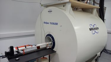
Sited directly next to the magnet room is an equipped surgery. Here, anaesthesia can be provided with animal physiology monitoring (breathing rate, ECG, blood pressure, body temperature). These monitoring methods can be reproduced inside the magnet for animal well-being.
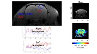
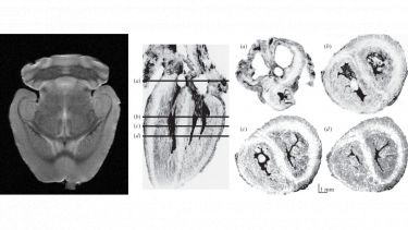
The scanner is routinely used in mouse and rat brain imaging at high resolution, typically voxel resolution is 100 μm but can be smaller if required. Images can be enhanced with gadolinium contrast agents, for example to highlight tissue lesions.
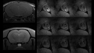
Rapid EPI imaging can be used to estimate cerebral blood flow for the assessment of disease progression. These experiments can be expanded to perfusion, for a marker of blood/brain barrier integrity. EPI imaging is often used in functional imaging (fMRI). Scanning whilst applying shorts period of stimuli can detect regional changes in brain activity.
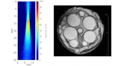
Working with the Department of Materials Science & Engineering, we have used the 7T scanner to study the behaviour of flowing fluids under stress. The enhanced sensitivity available at 7T field strength means that high resolution velocity maps can be produced. MRI is well suited to material studies, including biomaterials such as silk.
9.4T NMR/MRI scanner
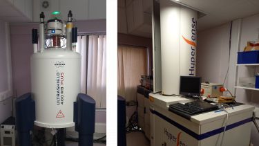
The 9.4T scanner is a dual-purpose instrument capable of both magnetic resonance imaging and high-resolution magnetic resonance spectroscopy. Equipped with an automatic sample changer, the scanner can perform high throughput liquid state NMR spectroscopy. It can also analyse biopsy tissue samples. In imaging mode, the combined field strength and gradient strength permits imaging with <50 μm resolution. The facility also houses a hyperpolarisation instrument (HyperSense) that can increase the sensitivity of NMR by many orders of magnitude. This can be used for measuring metabolism and reaction kinetics in real time.
High Resolution Spectroscopy
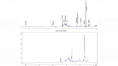
For high resolution NMR spectroscopy, the scanner is equipped with a multinuclear probe capable of observing all of the standard nuclei, e.g. 1H, 13C, 31P and many more (frequency range 1H–15N). It has been used extensively to perform metabolomics studies of cells and biofluids. An automatic sample changer allows 16 samples to be scan without user input for high throughput scanning and overnight/weekend data acquisition. The scanner also has a high-resolution MAS probe that can be used to analyse tissue samples and biopsies.
Microimaging
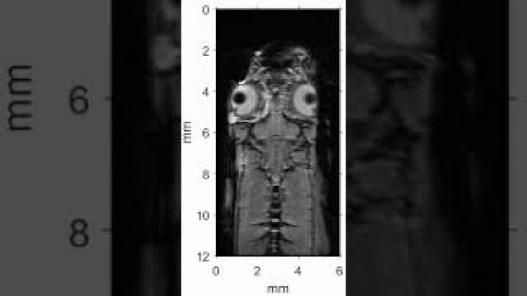
The ultra-shield 9.4T magnet allows the scanner to produce high quality images with improved signal to noise ratio (SNR). This can shorten scanning time to reduce the duration of animal experiments. The system is equipped with a powerful gradient set (1500 mT/m) allowing micrometre resolution imaging as small 15 μm in plane voxel size.
Zebrafish have become a ubiquitous animal model for studying a range of diseases and conditions. Typically, these studies are conducted in transparent juvenal fish where optical imaging techniques can be used. However, this is more difficult in adult zebrafish, restricting their use in longitudinal studies. Developed within the preclinical MRI facility, we have the capability to image live zebrafish. The animal can be scanned at high resolution (In plane, 50x50 μm, 200 μm slice thickness) with easy loading and full recovery to facilitate longitudinal studies.
Hyperpolarisation
The HyperSense instrument uses dissolution dynamic nuclear polarisation (dDNP) to increase the sensitivity of magnetic resonance spectroscopy (see our Hyperpolarisation webpage for the physics behind the technique).
To date the method has been extensively used in studying pyruvate metabolism, a key molecule in cellular energy generation. The experiment involves administering a bolus of hyperpolarised pyruvate to an animal or cells and rapidly measuring the 13C MRS spectrum in real time. As pyruvate is metabolised, daughter products such as lactate and bicarbonate can be detected. The rate of formation of these products can inform on the relative importance of glycolytic metabolism (lactate) and oxidative phosphorylation (bicarbonate). High levels of lactate are a marker of cancer, and when combined with imaging DNP can detect tumour cells and their response to response to treatment. The technique has been applied to other target organs, including the heart, kidney and brain.

Equipment
The equipment the facility has available is listed below.
|
7T MRI (Alfred Denny Building G Floor) |
Horizontal Bore Bruker Biospec (70/30S)
|
|
9.4T MRI/S (Jessop Wing, Level 4) |
Imaging: Vertical Bore Bruker MRI/S system
Spectroscopy:
|
| Other |
13C Hyperpolarisation - HyperSense:
|