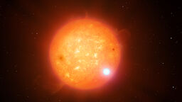
Life Under the Microscope: Building a centre of excellence for biological imaging technology

Physicists and biologists in Imagine: Imaging Life, a research group at the University of Sheffield, use and develop the most cutting edge microscopes to get an in depth look at our cells, at bacteria, and other microscopic entities

Microscope made using lasers

Many diseases are on the rise. Due to an increase in the number of antibiotic resistant strains of bacteria such as MRSA, the risk of untreatable infection is growing. And, despite many billions of pounds spent fighting it, cancer remains one of the major killers.

In order to cure disease, researchers first seek to understand how that disease affects cells. To do this, they must understand the structure of those cells and how a disease changes that structure.

However, cells and bacteria are invisible to the naked eye, and viruses are even smaller – too small for conventional microscopes to see.

Fortunately, academics in Imagine: Imaging Life, a research group at the University of Sheffield, use and develop the most cutting edge microscopes to get an in depth look at our cells, at bacteria, and other microscopic entities. Their data might ultimately be used to develop treatments for diseases such as MRSA and cancer.

Light it up: Optical microscopy

Dr Ashley Cadby from the Department of Physics and Astronomy at Sheffield, primarily works with different types of light microscopy, including super-resolution light microscopy, which has a resolution 10 times higher than the standard optical diffraction limit. This gives Ash and his team the ability to take images of living tissues at a resolution of about 20 nanometres (four times smaller than a flu virus).

Ash's main area of interest is in creating new microscopes and cameras, and has helped develop a camera that can collect data that has never been collected before.

He's also developed a technique that allows him to control how much light is put in and out of a single microscope, taking it from low-light to super-resolution microscopy in a millisecond.

"We’ve got to the point now where we can pick individual bacteria and study them with totally different microscopy systems, even though they are on the same microscope," said Ash. "It’s basically a transformer; we just need a cool name for it like Bumblebee. Right now it's called the Cairnfocal."

See the images captured on Flickr

'Feeling' sample surfaces: Atomic force microscopy

Atomic force microscopy (AFM) is a type of microscopy that forms an image by "feeling" the surface of a sample with a very sharp point. It allows Professor of Physics, Jamie Hobbs, and his team to obtain images down to the molecular scale, and can image "living" samples in their natural, physiological environment.


We are now able to start linking how molecules are organised to the ultimate properties that that gives a biomaterial, and through that to how it controls the biological function.

Professor Jamie Hobbs

Imagine researcher


"One of the unique capabilities of AFM is that it can measure the mechanical properties of a sample at high resolution, so we are now able to start linking how molecules are organised to the ultimate properties that that gives a biomaterial, and through that to how it controls the biological function," said Jamie.

Cells and molecules in 3D: Cryo-electron microscopy

The cryo-electron microscopy (cryo-EM) microscope has the highest overall resolution of the three. By using electrons, it gets past the light diffraction limit and can take pictures with detail visible down to about three angstroms (0.3nm). For reference, the size of an atom is about one angstrom.

This allows groups in Sheffield, including Professor of Structural Biology Per Bullough and his team to take three dimensional images, which give them insights into the physical characteristics of cells at a near atomic level, and provide a far better understanding of how molecules in cells interact with one another.

"To understand how cells work, we need to understand the sheer complexity of them, and to understand that we need to know what they look like in all their fine, three dimensional detail." said Per. "It’s a great challenge as cells have a lot of components and they are very dynamic."

The cryo-EM technology is so ground-breaking that three of the scientists behind it won the 2017 Nobel Prize in Chemistry.

One of them, Richard Henderson, was Per’s PhD supervisor. He gave the annual Krebs Lecture in 2018 to coincide with the unveiling of the University of Sheffield's new Arctica cryo-EM facility.

Nobel Prize winner Dr Richard Henderson with his former PhD student Professor Per Bullough  at the launch of the University of Sheffield's new cryo-electron microscopy facility.
Nobel Prize winner Dr Richard Henderson, left, with his former PhD student Professor Per Bullough, third from left, at the launch of the University of Sheffield's new cryo-electron microscopy facility

Blurring scientific boundaries

Though both Jamie and Ash are part of the Department of Physics and Astronomy, their research heavily overlaps with biology, as they work with biologists to tackle biological problems and where necessary develop microscopy techniques to solve them.


To understand how cells work, we need to understand the sheer complexity of them, and to understand that we need to know what they look like in all their fine, three dimensional detail.

Professor Per Bullough

Imagine researcher


Jamie and his group particularly focus on the bacterial cell wall and the action of antibiotics, on breast cancer metastasis (its spread from one site to another, secondary site), and on the plant cell wall and plant morphogenesis (how they take shape). Recently, they have also started exploring DNA-protein interactions.

So far, some of the discoveries made during this research have literally rewritten the textbook when it comes to models of the bacterial cell wall, with an image taken by Jamie's team appearing in Brock Biology of Microorganisms.

Per's main area of interest is in seeing how molecules come together and interact with each other. This research involves using cryo-EM to see the physical processes that occur during these interactions. He also works closely with Ash and Jamie.

Together, the three researchers, along with Professor of Molecular Biology Simon Foster, are the coordinators of Imagine: Imaging Life.

Imagine brings together all three types of microscopy and allows academics to share their knowledge and facilities with one another, resulting in them getting a far fuller understanding of how living systems work.

"Getting all the technologies running at what is truly the cutting edge has taken a bit of time, but we are now really starting to bring the different ways of imaging to bear on difficult and pressing problems," said Jamie.

"The cancer example is a good one. Locally measuring mechanics at the length scale that is felt by individual cancer cells is only really possible with AFM. But if we didn’t have the expertise in optical microscopy to hand we would be very limited in what we could do with that information – it would lack context."

Imagine employs a number of technicians and PhD students who do their own individual projects using the microscopes.

The opportunity to use Imagine's facilities and data is also available to undergraduates, who can work in the labs for their final year projects or as part of the Sheffield Undergraduate Research Experience summer placement scheme. And a new postgraduate course MSc Biological Imaging, has been created to teach postgraduates how to use all three technologies and complete a microscopy research project.

"Honestly," said Ash, "the best thing about Imagine is the students. Imagine not only gives students a chance to do a PhD or MSc, but also lets them see how their work fits in with the wider world of science. This means we end up with students who are not just good with one technique – they’re good at biology and microscopy in general."

Large crowd of Imagine researchers stood on steps at the project's 2017 launch event.
Imagine researchers at the project's 2017 launch event.

Imagine's work has already had an impact on the study of pathogens. For example, a recent collaboration between Per and Dr Robert Fagan in the Department of Molecular Biology and Biotechnology is shedding light on the structures of potential targets for antimicrobials in C. difficile, a common and serious hospital infection. Though they don’t have any firm answers yet, Cryo-EM will bring new insights.

The future of microscopy

Microscopy has come on leaps and bounds in the last few years, and things are only getting started. With three cutting edge types of microscopy all under one banner, the possibility for collaboration grows by the day. And as the technology continues to improve, new discoveries will become more and more frequent. However, this progress will not continue without keen, intelligent new researchers entering the field.


The best thing about Imagine is the students. We end up with students who are not just good with one technique – they’re good at biology and microscopy in general.

Dr Ashley Cadby

Imagine researcher


"We need collaborators – people who want to work with people to get science done," said Ash.

"We need people to build microscopes, to collect the data, and to analyse that data. It's collaboration that will lead to advances in things like cancer and antimicrobial resistance research."

Though the discoveries made so far have been incredible, researchers have only really begun to scratch the surface of what is possible when all three microscopy methods are combined. Looking ahead, Per, Ash, and Jamie are all very hopeful for the future.

"In the coming decades, we’ll have a picture of an entire cell, and understand in fine detail all their components and how they interact," said Per. "That’s what the next generation of researchers has to look forward to."

REF 2021 illustration showing University building and subject areas

Research Excellence Framework 2021

We have been rated 1st in the UK in terms of the quality of our research. In the latest REF, 100 per cent of research and impact from our department has been classed as world-leading or internationally excellent.

Four students laughing while sat at a bench, outside the Students' Union

International Merit Scholarships

We offer a generous package of financial support for international students including 75 undergraduate scholarships worth £10,000 towards the annual tuition fee and 125 postgraduate taught scholarships worth £5,000 towards the tuition fee. Applications are now open for existing offer holders.