Confocal Microscopy
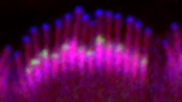
Confocal microscopy is a powerful imaging technique that provides high-resolution, high-contrast images by using point illumination and optical sectioning to eliminate out-of-focus light. This allows for detailed visualisation of fluorescently labeled structures within thick tissue samples.
In our research, we use confocal microscopy to image specific regions of the auditory system that have been labeled with fluorescent markers targeting particular cell types and proteins. This approach enables us to precisely localise and quantify cellular and molecular changes associated with different forms of hearing impairment. By visualising how the structure and distribution of key components are altered under various conditions, confocal microscopy helps us uncover the biological processes underlying hearing loss and assess the impact of experimental interventions.