
Morphology
To understand how sound is processed and the basis of hearing loss and deafness, we aim to identify the location, appearance and interaction between the key components of the auditory system. We do this by using powerful microscopes that can visualise structures down to a 100,000 times smaller than the width of a hair. Click here to find out more.
To investigate the morphology of the cochlea and sensory cells, we use a combination of confocal and electron microscopes available at Sheffield. These approaches enable us to study how sensory cells connect to auditory neurons and the structures responsible for transducing sounds. Furthermore, we are working on understanding how these structures are affected in mouse models of hearing loss and deafness.
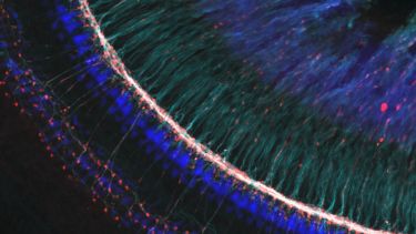
This is an example image taken from a confocal microscope. It labels auditory nerve fibres (cyan and magenta) and hair cells (blue)
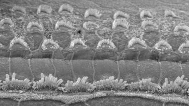
Scanning electron micrograph showing a single row of inner hair cells at the bottom of the image and three rows of outer hair cells at the top.