MR hardware engineering
We have a strong pedigree in MRI hardware engineering, including pioneering work on RF coil, noble gas nuclear hyperpolarisation, and low field MRI hardware development.

RF coils
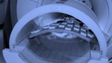
The radiofrequency (RF) coil is the closest instrument to the subject under examination in MRI. It is the final component in the transmit chain—determining the uniformity of flip angle and power efficiency—and the first component in the receive chain—determining the image quality. The RF coil is a series resonant circuit, wherein the inductor is a distributed component capable of generating and detecting near-field oscillating magnetic fields. During transmission, the RF coil generates an oscillating magnetic field orthogonal to the static magnetic field, tipping the bulk magnetisation of the sample and the receiver RF coil detects this signal before it decays.
The oscillating magnetic field interacts with the bulk magnetisation of the sample only when the frequency is the same as the Larmor frequency. Different nuclei have discrete Larmor frequencies according to their gyromagnetic ratios:
| Nucleus | Gyromagnetic Ratio (MHz / T) |
|---|---|
| 1H | 42.6 |
| 3He | -32.4 |
| 13C | 10.7 |
| 19F | 40.1 |
| 129Xe | -11.8 |
Detection of each nuclei requires dedicated multi-tuned RF coils, or sets of coils that resonate at multiple frequencies, thereby imposing RF engineering challenges to enable multinuclear MR capabilities whilst electrically isolating resonant circuits and minimising losses. Resolving these challenges by designing novel RF circuits, components and multinuclear RF coils is the primary focus of engineering research at the POLARIS group - with an intent to apply such technology in the clinical setting.
X-nuclei RF coils
There are few commercially available RF coils for X-nuclear (non-1H) applications, especially when the anatomy of interest is specific, for example brain, lungs, breast and kidneys. In addition, x-nuclei typically resonate at much lower frequencies compared to 1H, which adds additional challenges to circumvent poor sample loading and filling factor. As such, X-nuclei RF coils often need to be developed in house for specific needs.
- Featured Example: 129Xe Brain coils
-
Two birdcage transmit-receive RF coils at 1.5 T and 3.0 T were developed for MR imaging and spectroscopy of inhaled hyperpolarised 129Xe in the brain. Using these RF coils, we were able to detect five distinct spectroscopic peaks in the human head for the first time.
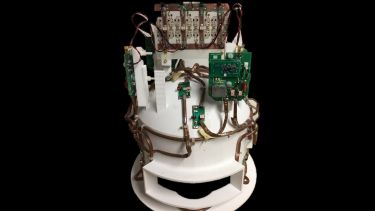
4 channel 129Xe brain coil Furthermore, a four channel receiver RF coil was developed in house with superior grade copper, and we were able to directly image inhaled hyperpolarised 129Xe in the human brain. We were also able to show that in a stroke patient the region affected by impaired gas-exchange in the brain is much larger than those seen by contemporary imaging methods.

Multinuclear RF Coils
There are many approaches to build multinuclear RF coils:
- a common resonant structure that resonates at multiple resonant frequencies
- switchable RF coils using actively-controlled switches
- multiple RF coils resonating at different resonance frequencies, and that are electrically isolated
- using meta-materials

At POLARIS, we have investigated the theory of trap circuits—passive parallel resonant circuits used in series with the RF coil—and we were able to establish an accurate mathematical formula to calculate the component values to build multinuclear RF coils with an arbitrary number of traps. We have demonstrated triple nuclear MR imaging of 129Xe, 3He and 1H, and dual nuclear diffusion imaging of 129Xe and 3He in a breath hold at the same lung inflation state. In addition, we have compared the use of micro-electro-mechanical-systems and PIN diodes for switchable RF 19F-1H coils. 19F is another emerging imaging agent for lung MRI, and the key engineering challenge is the proximity of the resonance frequency to that of protons. We recently proposed an 8 channel distributed transmitter-receiver RF coil with 6 additional receive only channels as the optimal solution for multinuclear imaging of 19F and 1H of the lungs.
People, Projects & Publications
-
People
-
-
Current Projects / Grants
-
We have ongoing projects in the development of:
- High-density RF coils for multinuclear MRI of the lungs, brain and kidney, e.g.
- 6 channel 129Xe brain RF coil for 3.0 T
- 8 channel dual-tuned 1H-129Xe RF coil for 1.5/3.0 T
- Novel circuit components for multinuclear MRI
Our work in MR hardware engineering is / has been supported by the National Institute for Health Research (NIHR), Medical Research Council (MRC), Global Challenges Research Fund (GCRF), BRC grant awards, a studentship from the Natural Sciences and Engineering Research Council of Canada (NSERC), University of Sheffield faculty scholarships, and collaborative funding from the Memorial Sloan Kettering Cancer Center (MSKCC). Current main grants include:
- 04/2015—03/2023. Expansion of state-of-the-art MR imaging infrastructure for pulmonary disease stratification: POLARIS. MRC - Resources and infrastructure Research Grant. PI: Wild
- 06/2016—05/2024. Xenon gas recycling project. Linde gases. PI: Wild
- High-density RF coils for multinuclear MRI of the lungs, brain and kidney, e.g.
-
Past Projects / Grants
-
- 11/2013—10/2018. Translating novel pulmonary MR imaging methods in to clinical practice. NIHR - Research Professorship. PI: Wild
- 10/2012—09/2016. Hyperpolarised 129Xe Magnetic Resonance Imaging Techniques for Assessment of Human Lung Function. MRC - CASE Industrial Studentship with GE Healthcare. PI: Wild
- 10/2006—10/2011. Enhancement of Sensitivity in the MRI of Hyperpolarised Noble Gases. EPSRC - Advanced Fellowship. PI: Wild
-
Collaborators
-
We work closely with the following MR industry partners:
- GE Healthcare
- Clinical MR Solutions
- Philips
- PulseTeq
- Voxelgrids
-
Key Publications
-
-
Maunder A, Rao M, Robb F & Wild JM (2020) An 8-element Tx/Rx array utilizing MEMS detuning combined with 6 Rx loops for 19F and 1H lung imaging at 1.5T. Magnetic Resonance in Medicine, 84(4), 2262-2277
-
Maunder A, Rao M, Robb F & Wild JM (2018) Comparison of MEMS switches and PIN diodes for switched dual tuned RF coils. Magnetic Resonance in Medicine, 80(4), 1746-1753
-
Rao M, Stewart NJ, Griffiths PD, Norquay G, Wild JM (2017) Imaging Human Brain Perfusion with Inhaled Hyperpolarized 129Xe MR Imaging. Radiology, 286(2)
-
Rao M, Stewart NJ, Norquay G, Griffiths PD & Wild JM (2016) High resolution spectroscopy and chemical shift imaging of hyperpolarized 129Xe dissolved in the human brain in vivo at 1.5 tesla. Magnetic Resonance in Medicine, 75(6), 2227-2234
-
Rao M & Wild J (2016) RF instrumentation for same-breath triple nuclear lung MR imaging of 1H and hyperpolarized 3He and 129Xe at 1.5T. Magnetic Resonance in Medicine, 75(4), 1841-1848
-
Rao M, Robb F & Wild JM (2015) Dedicated receiver array coil for 1H lung imaging with same-breath acquisition of hyperpolarized 3He and 129Xe gas. Magnetic Resonance in Medicine, 74(1), 291-299
-