

- New project entitled ‘Mapping the Brain’ is focusing on new ways to scan and map the brain
- The project could lead to more efficient brain scans and could speed up research into aspects of brain health such as diseases and sensory perception
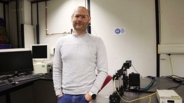
- Dr. Andrew Maiden from the University of Sheffield is collaborating with Professor Andreas Schaefer at the Francis Crick Institute for the project
A new £200,000 grant from the Biotechnology and Biological Sciences Research Council (BBRSC) awarded to the University of Sheffield, in collaboration with the Francis Crick Institute in London, could help us better understand the inner workings of our brains.
The project, called ‘Mapping the Brain’, is part of the BBSRC’s ‘Basic Technologies in Sensing and Imaging’ programme. Dr. Andrew Maiden, Senior Lecturer in Computational Imaging in Sheffield’s Department of Electronic and Electrical Engineering, and Professor Andreas Schaefer, Professor of Neuroscience at University College London and Principal Group Leader at the Francis Crick Institute, are focusing on new ways we might scan and map our brains to better understand everything, from diseases to our sensory perception.
The work sits within a field known as connectomics, which centres around the tracing of the interconnections between neurons. These synapses, which connect neurons together, are only nanometres in diameter, but they can stretch out over several millimetres. The investigation of how these neurons work together is one of the top priorities for neuroscientists, as the answers could be the key to unlocking the secrets of the human brain.
What my collaborator, Professor Andreas Schaefer at the Crick, wants to see is how these nano-scale tendrils connect cells with each other over millimetre-cubed volumes. That's difficult, because first of all you have to see the tendrils, which are really, really thin, but then you also have to map out these huge volumes.
Dr. Andrew Maiden
Senior Lecturer in Computational Imaging at the University of Sheffield
The current approach for brain mapping, currently carried out at the Crick, is for a cube of brain tissue a few hundred microns thick to be analysed by researchers. This is done by shaving extremely thin slices from the cube for imaging using an electron microscope. The process is carried out repeatedly to build up a map of the tissue, slice by slice. There are, however, challenges with this approach.
One of the problems with slicing brain tissue in this way is that many sections will get lost or damaged when collecting thousands of slices, which causes serious issues with reliability and scalability. The other issue is that even though researchers currently only use a small volume in each scan, it still takes a huge amount of time to collect the results - for high image resolution scans, the machine mapping may have been running for months, or even years to capture a volume of only a fraction of a cubic millimetre.
This new collaborative project between Dr. Maiden and Professor Schaefer will integrate an X-ray method to complement and target the existing process of electron microscopy (wherein each slide of the brain is examined one at a time). The outcome will not be quite as high resolution as the electron microscope process would provide, but will allow the researchers to look at much bigger pieces of the brain, without physically cutting it. This process is known as multi-slice ptychography.
“The idea is that you get this slightly worse resolution but significantly larger-volume map of the brain tissue, which will then allow our colleagues at the Crick to say ‘oh, well that bit is interesting, let’s investigate that further, we'll look at that with our electron microscope.’ So it's a correlative technique. We image using the X-ray method over a big volume, so then you can target where you want to get the extra detail from the electron microscope.” Dr. Maiden said of the aims for the 'Mapping the Brain' project.
Dr. Andrew Maiden is working with the Diamond Light Source at Harwell in Oxfordshire to develop this new technique.
Expanding on the techniques involved in the research, Dr. Maiden said: “The high energy X-rays at Diamond will allow us to penetrate full cubic millimetre brain tissue samples and we hope eventually this will enable us to image this tissue in three dimensions at nano-scale resolutions. Our method, which is called ptychography, measures the diffraction patterns generated by the X-ray beam as it passes through the brain tissue, then uses algorithms originally developed in Sheffield by myself and Professor Rodenberg to process the diffraction data and produce a 3D image.”
Professor John Rodenberg, Emeritus Professor in the Department of Electronic and Electrical Engineering at the University of Sheffield, helped to pioneer the X-ray technique that is used in this research in the early days of its development. Professor Rodenberg is a Fellow of the Royal Society, and it was his work on this that originally brought Dr. Maiden to the University of Sheffield.
The hope for the research being carried out within the BBSRC supported programme is that by mapping these brain connections we can better understand neuro-degenerative diseases, such as Parkinson's. It could also lead us to better understand the human senses - the olfactory system, which is the sensory system used for sense of smell, is a particular interest of Dr Maiden’s colleague at the Francis Crick Institute. If researchers are able to better understand how the brain connects neurons together, and if they are able to do so at a far faster rate, then aspects of brain health such as disease and sensory perception could be understood at a deeper level far sooner.